Time
Time is a universal governing force in our world, the effects of which may be readily observed in the realm of biology and medicine. After all, time is the determining factor in our own mortality, a reality we are reminded of each time a loved one or beloved family pet dies. Indeed, life on earth is made possible only by the death of other plants and animals, linking the two in a continuous cycle of ends and beginnings.
Time is an abstract yet basic concept that defines the existence of every human being and the world in which they live. Everything we experience – from music and the formation of relationships to embryologic growth and the disease process – occurs in time, making it an inescapable and defining element of our existence. The study of medicine, too, is largely concerned with the passage of time, as a great many physiological cycles and developmental and aging processes occur from second to second, hour to hour, month to month, and year to year. The images presented below are intended to convey the centrality of time in the study of medicine and biological processes.
Life time – 6 Images
9 months and then a Lifetime |
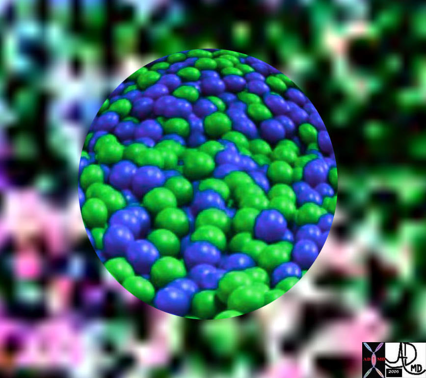
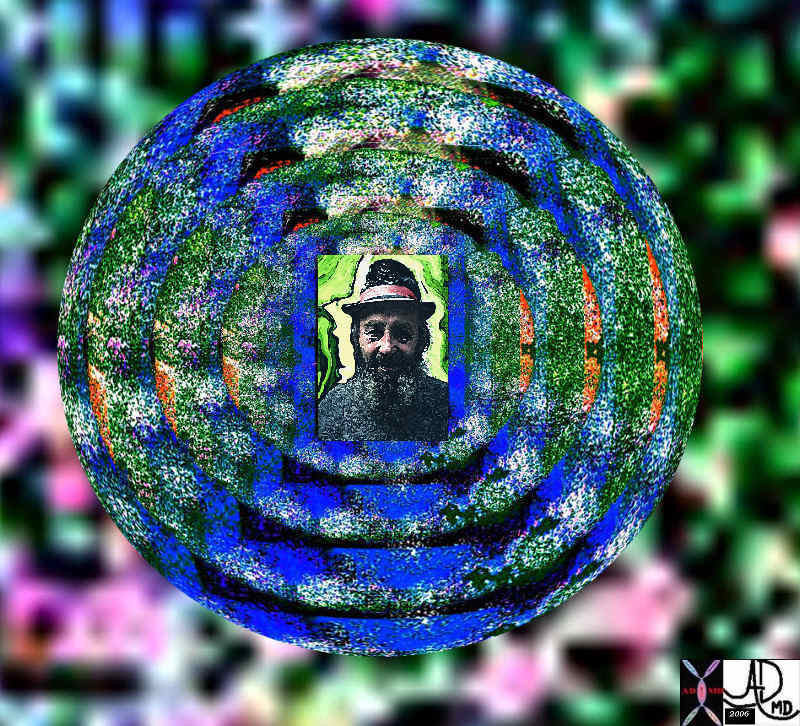
| These 6 images look at time from a more embryonic perspective, from the time of conception (top left) where sperm meets egg, through the multiplication of cells as the blastocyst evolves, through fetal development as the primitive tissues evove into a fetus attached by an umbilical cord (top right), a fetus flexed in the amniotic cavity with fully formed organs, a boy in full extension in his stride, and an old man who has been through a number of life cyles at the latter end of his life cycle.
13275b01i01e12u08 Davidoff oneness |


Time in Structure
All the structures of the body will theoretically have a time zero at the moment the sperm fertilizes the egg. From then on structures will grow, mature, will be subject to the joys and wraths of time, and hence will change, and then all will die.
There are both linear and cyclical elements that relate to the way structure relates to time. Some structures will grow in size with time, such as the long bones of the body, and some structures like the ovary will undergo cyclical changes with time. These will both be explored in the discussions below.
Growth
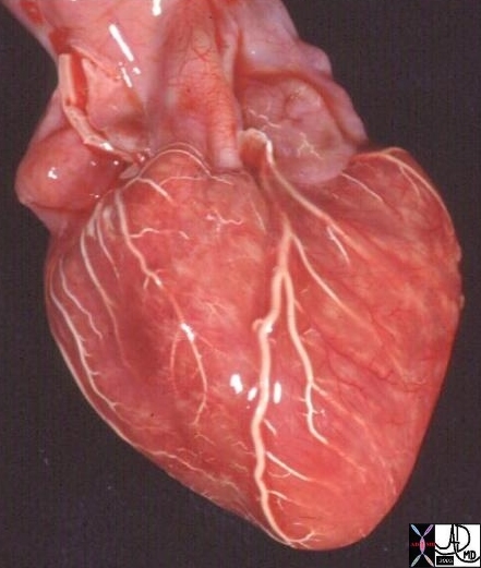
The growth phase for some tissues is phenomenally fast. Fetal brain tissue for example is said to grow at 250,000 nerve cells per minute. The heart starts out as a microscopic blob of mesoderm, a primitive embryonic tissue. Deeply embedded in the genetic makeup of the human form, there are codes that will direct the mesodermal blob to develop from a straight tube to a complex beating pump in 5-6 weeks. At that time it will have the ability to connect to all parts of the body which at the time is a mere 3-4 mm long. By this time it is a 4 chambered mature organ. From then on its growth is mostly a change in size in order to adapt to the increasing size of the body.
  Heart Development Heart Development
By 5-6 weeks the mature structure is developed |
| The heart starts out as a blob of mesoderm, and progresses through a straight tube phase, loops (usually to the right) twists, septates, and becomes a 4 chambered structure by 5-6 weeks.
01477b03 heart cardiac straight tube primitive heart embryology Davidoff art drawing Davidoff MD 15009c cardiac heart coronary artery normal anatomy grosspathology LAD diagonal artery acute marginal artery conal artery arc of Vieussens Courtesy Ashley Davidoff MD |
Some bones on the other hand, continue to gow for many years, and will have unfused growth plates till the early twenties. The long bones of the body, like the femur and humerus are examples. The growth plates (aka epiphyseal plates ) at either end of the long bones are made of cartilage. They continue to evolve and grow by mitosis until they finally ossify at maturity. While growing, the cartilage cells in the region called chondrocytes, age and degenerate. Osteoblasts move in and ossify the matrix to form bone. This process continues throughout childhood and the adolescent years until the cartilage growth slows and finally stops. When cartilage growth ceases, usually in the early twenties, the epiphyseal plate completely ossifies so that only a thin epiphyseal line remains and the bones can no longer grow in length. Bone growth is under the influence of growth hormone from the anterior pituitary gland and sex hormones from the ovaries and testes.
The humerus serves as a skeletal guide reflecting maturation from adolescence into adulthood. The distal growth plate is the first of the long bones to unite, while the proximal growth plate is the last of the growth centres to unite. This occurs at about 18-20 years. The growth plates of the iliac crests fuse a little later (early twenties) while the sutures of the vault may close beyond that age.
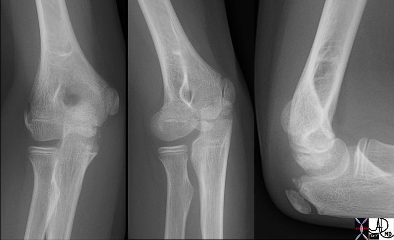
Epiphyses at the elbow of an 11 year old |
| The X-rays of an 11 year old child reveal unfused ossification centers of the distal humerus, proximal radius, and ulna. They appear as unfused “fragments” but are really cartilagenous areas that progressively ossify during growth until they ossify completely and fuse at maturity. The distal humerus is the first of the long bones to unite.
73590c01.800 11 years old with ossification centers elbow humerus radius ulna bone normal anatomy ossification centers 6 ossification centers around the elbow joint. appear at different ages or fuse. the ossification centers always appear in a specific sequence. mnemonic of the order of appearance of the individual ossification centers is C-R-I-T-O-E: Capitellum, Radial head, Internal (medial) epicondyle, Trochlea, Olecranon, External (lateral) epicondyle. as a general guide, remember 1-3-5-7-9-11 years. |
Even though bones stop growing in length in early adulthood, they can continue to increase in thickness or diameter throughout life in response to stress from increased muscle activity or to weight. The increase in diameter is called appositional growth. Osteoblasts in the periosteum form compact bone around the external bone surface. At the same time, osteoclasts in the endosteum break down bone on the internal bone surface, around the medullary cavity. These two processes together increase the diameter of the bone and, at the same time, keep the bone from becoming excessively heavy and bulky.
 Young and Aging Young and Aging |
| Normal changes in the skin over time as a result of loss of elasticity of the tissue.
49609c03 abdomen health disease young old time cycle elastic elasticity order disorder Davidoff MD 49609c02 |
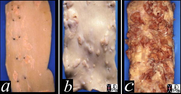
In the cardiovascular system early “normal” atherosclerotic changes are seen as early as 18 years and the macroscopic result is is called fatty streaking. This is due to normal wear and tear and causes no harm or disease at this time. It is purely an early “wear and tear” on a structure that is undergoing expansion and relaxation about 60 times a minute, (conservative estimate since during sleep heart rate is slower), 3,600 per hour, 217,728,000 per year, 4,354,560,000 after 20years, and 15 billion by 70 years. Thus wear and tear changes should not be unexpected and are considered “normal”. However in the presence of other factors like hypertension, dietary and behavioral indiscretions, or genetic factors, the degenerative process is accelerated and diseases such as aneurysms and obstructions can occur.
 Atherosclerosis Atherosclerosis |
| This image shows three pathological specimens of the aorta. In the first image minimally raised fatty streaks are noted. (a) This is a normal “wear and tear” finding and can be seen as early as 18-20 years. In image b, the fibrous capsule causes raised fibrofatty nodules. This is also a normal appearance, and would be seen in the aorta of a 60-70 year old. In c, there is severe atherosclerosis, more than expected for wear and tear, and this is pathological. There has been rupture of the plaques, resulting in friable atheromatous plaques.
Courtesy Henri Cuenoud MD 13420c |
Life Cycles
Some biological units like the patelet will complete a life cycle in a mere 10 days. The red cells have a life span of only 120 days after which time they are removed from the circulation, broken down by the spleen and parts are either excreted or reused in some form.
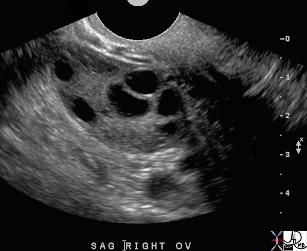
All the ova of an individual (400,000) will be “born” with the ovary and will remain latent till female maturation. Only about 450-500 will reach maturity during the reproductive life of the female. Each month during the years of reproductive capability, one ovum will reach maturity and will be deployed for potential fertilzation. The ovum will last 12-24 hours and will either end its cycle if not fertilized, or go onto an exciting roller coaster ride for the next 70 years or so if it is fertilized. This process continues till menopause.
 Follicles in a Reproductive female – Cyclical Phases Follicles in a Reproductive female – Cyclical Phases |
| This ultrasound image shows 5 developing follicles in the right ovary that are all less than 1cms at this stage. (black cystic structures) One of these will progress to ovulation and will release its ovum when the follicle becomes about 2cms.
71689 ovary follicles normal anatomy function physiology TCV Applied Biology Cycle time USscan Davidoff MD |
Time in Biologic Function
It has often astounded me throughout my medical education how many hours a premedical or medical student may put into gaining mastery of a process that occurs in a mere matter of seconds. One example of this is the action potential, the impulse that arises from the rapidly coordinated opening and closing of sodium and potassium channels in the neural membrane. This event, which occurs in fraction of a second, is a defining one in human function, as it is the basis of the nervous system and all sensory and motor control. The summation of action potentials to convey more coordinated impulses and more detailed information to and from the CNS is also critical to proper physiologic functioning and, incidentally, serves as yet another example of units to unity.
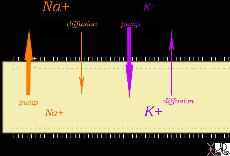 Sodium Pump and Potassium Diffusion Process in the Creation of an Electrical Path fo the Transmission of Neural Impulses Sodium Pump and Potassium Diffusion Process in the Creation of an Electrical Path fo the Transmission of Neural Impulses |
| 72045b04.800 nerve conduction force electricity electric force positive force negative force sodium pump Na pump Patassium pump K+ pump diffusion conduction of impulses Davidoff drawing Davidoff art Davidoff MD |
Perhaps one of the most important ways in which action potentials manifest themselves is in the regular, coordinated beating of the heart. Equipped with its own pacemaker, the SA node, the heart experiences regular contractions of its atria and ventricles about every second such that blood may be continually pumped through the body. The rapidity with which the SA node issues electric impulses is, in turn, responsible for the second-to-second changes in the shape and width of blood vessels, which expand and contract as blood pulses through them. However, the cardiovascular system is but one illustration of the complex functions that may be achieved along the order of seconds. Countless chemical reactions occur with astounding speed at such critical locations as the active sites of enzymes, cellular organelles, and the interiors of each organ, such as the liver. The images below serve as illustrations of some of the aforementioned processes.
Changes per Second
 EKG – Electrical Impulses EKG – Electrical Impulses |
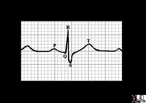
| The EKG reflects the electrical activity of one heartbeat and consusts of a p wave, a QRS complex, and a t wave. This event takes place in about 3/4 of a second in this instance.
71679 heart cardiac electrical activity electricity force p wave QRS complex STsegment T wave normal EKG Laboratory Davidoff MD |
In the above example the EKG shows the time over which the electrical activity occurs through one cardicac beat. Each small square is .04seconds or 40 miiliseconds. The p wave is the depolarisation process of the atria and in this instance it takes just under 3 squares or .12seconds or about 120 milliseconds. (normal adult (120-200msecs) The QRS complex is the depolarisation process of the ventricles and it takes about 80 milliseconds. (normal adult (60-100 milliseconds) The ST segment is an isoelectric period and takes about 320 milliseconds. The T wave is a repolarization process. The end of the T wave to the beginning of the p wave and a new cycle is about 200 milliseconds in this instance. Thus with a person at rest, the total cycle is about 750 milliseconds. The biochemical events that bring this about are occurring in nanoseconds while the electrical manifestations of depolarisation and repolarisation are measured in subsecond time- ie milliseconds.
Of course, proper physiologic function is not defined solely by the rapid activity one sees in chemical reactions and action potentials. Many processes, such as mitosis and meiosis occur in a matter of minutes and hours, depending on the organism. Certain bacteria, for instance, are capable of doubling and re-doubling their population in hours under ideal conditions, allowing for the rapid rise of infections in humans and other organisms. Cellular division is also essential in humans, where it occurs more slowly and underlies the processes of healing, tissue regeneration, and gametogenesis. Even respiration, another fundamental physiologic function, may be tracked according to the volume of air inspired over time, as illustrated below.
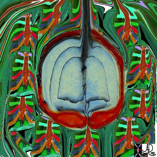 Time and the Respiratory System Time and the Respiratory System |
| This diagram represents a single cycle of respiration. The lungs expand on inspiration. At rest this takes about 2 seconds. They return to thir base line relaxed stae with expiration which takes about 3 seconds.
42530b05b09b14 Davidoff art |
Biological cycles are also intrinsically tied to the passage of minutes and hours, as they are defined by the occurrence of chemical or electrical events in regular intervals of time. For instance, ovulation and the menstrual cycle involve the precise and predictable adjustment of hormone levels over a certain number of days, such that any female experiences regularity in her reproductive cycle. In this way, the varied implications of biology as a function of time become clear.
Thus, the demise of a red blood cell or the progression from embryo to child to adult are characterized may be identified as a key player. In this way, it becomes clear that biology is inextricably bound to the passage of time.
Space
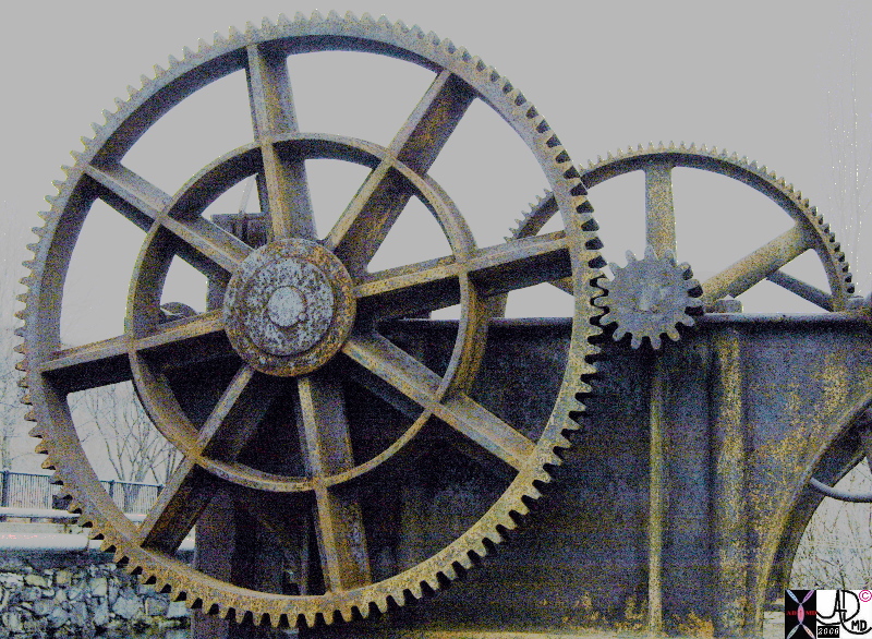
Space is a fundamental part of the universe. There are both scientific and philosophical concepts making a single definition difficult, but in the context of the Navigators Guide to the Medical Universe, it relates to the position in which matter exists, both as an absolute but usually in relation to other structures. In biology and the world within which we live, the positioning in space of one structure in relation to others is critical in the function of biological units. We also see the impotrance of positioning in non biological systems as demonstrated in the example below of cogs in the wheels of a mechanical tool. The positioning of the cogs of one wheel have to coincide with the cogs of the other in order for the turning of one to effect the turning of the other, and in the end for the functioning of that unit to occur.
|
Positioning of the Cogs is Essential to The Workings of the Whole System |
|
In this instance the cogs of the wheel in space are perfectly aligned with the cogs of the second wheel and so theoretically this machine as far as positioning, is ready to function. 86627.8 cogs in a wheel precise position Davidoff art Davidoff photography Lowell Massachusetts Remnants of days gone by copyright 2008 |

Cogs in Disorder – |
|
In this instance the cogs of these wheels are malpositioned as they lie in disarray on the ground and are thertefore far from being a functional unit. 89051p 89049p cactus gears junk discarded not functioning disorder Disney World Magic Kingdom Davidoff photography |
In the context of everyday living, space is usually a reference to the universe at large or to one’s “personal space.” With respect to biologic units, however, space refers to how cells, tissues, and organs are positioned within the body. While these two frameworks for understanding space may seem quite different they are actually very easily relatable. For instance, it is commonly understood that violation of one’s personal space is a source of considerable discomfort and tension, which one usually attempts to escape.

Did the Photographer Impinge on Her Space? |
|
This little girl was playing in the park and the approach of the photographer was sufficient for her to react with this expression which in effect says “Get out of my face and my space!” 02716p.8 girl in a park with doll anger protection space impingement Boston Massachusetts Davidoff photography Courtesy Ashley Davidoff MD Davidoff photography people copyright 2008 |
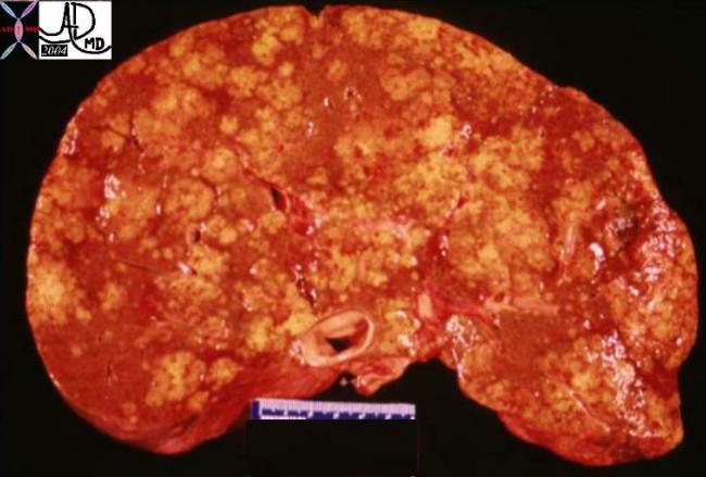
Organs, too, are negatively affected by encroachments upon their space by other factors – be they tumors, external pressure, or other abnormalities. One of the reasons that canceris such a devastating disease, is that it takes over the space occupied by normal functioning tissue. The liver is known as the metabolic warehouse of the body, and it is involved in many metabolic activities of the body. In the example shown below, a normal liver and a liver overwhelmed with cancer is shown. The cancerous components, not only occupy space, but they distort, and compress and destroy normal tissue and thus the liver is prevented from performing its metabolic functions.
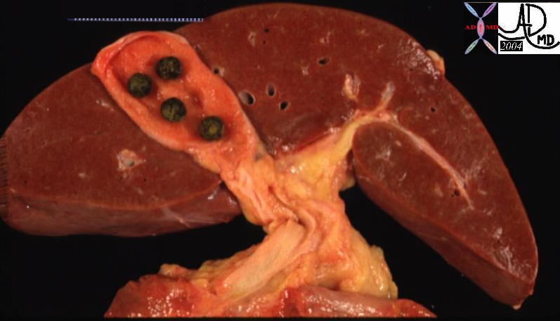 
Normal and Diffuse Metastases |
|
The left image is a anatomical specimen of a normal liver, though the gallbladder does contain 4 stones. In the specimen on the right the liver is overwhelmed with yellow cancer deposits that originated from a cancer in the duodenum. The cancer is only concerned about its survival and plays no part in contributing positively to the normal required processes of the body. By occupying the space of the normal liver it inhibits the function of the liver and also destroys the tissue. 13456 liver normal anatomy gallbladder stones cholelithiasis grosspathology 02635 metatstases to the liver from primary duodenal carcinoma |
A heart under increased pressure cannot pump as well. Pericardial tamponade is a life threatening condition in which the pericardium fills rapidly with a mere 100ccs of fluid . Tension pneumothorax is another life threatening condition in which air rapidly accumulates in the pleural space and encroaches on the the lungs and cardiovascular system. The examples of life threatening space occupation by disease are countless. It is clear that the positioning of structures relative to each other is critical to optimizing their functions, and maintaining viability.
Also crucial to the functioning of organs is the precise organization of their constituent cells and tissues. To illustrate this point think of each organ, and more specifically the glands in this instance, as a factory that is constantly at work to assemble small, simple raw materials into larger, more complex products. Once synthesized these products must be transported via tubular systems to target sites where they may be stored or put to work immediately. However, this assembly and delivery of products may only occur with optimal efficiency and fluidity if every screw, gear, and conveyer belt is in place. In the example below, the “factory line of the liver is demonstrated. For instance, the channeling of protein, carbohydrates, and fats into the liver from the portal vein only occurs properly if the hepatic cells are optimally arranged to receive them. Similarly, more complex products like enzymes and hormones may only be produced and transported from the liver if the cells and bile duct are properly positioned within the organ. A trash collecting system manned by the Kupffer cells lies at the edge of the sinusoids is also present to filter noxious material. The sinusoids represent the microvascular system that bathes the cells – almost like a river alongside which the cells are positioned to enable the receipt and transport of necessary products.
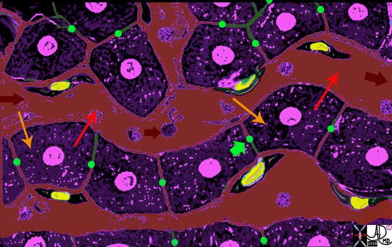
Positioning of the Liver Cell Around the Sinusoids |
|
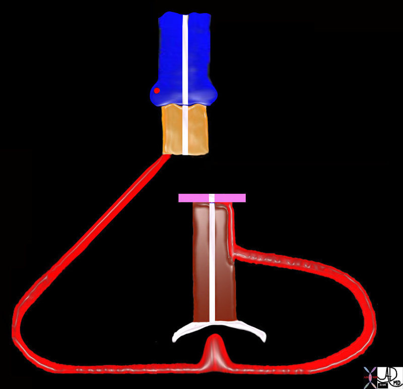
The liver cells (purple with central pink nucleus) are arranged in cords along the sinusoid (maroon) which is the smallest blood vessel of the liver. The portal vein brings metabolic products from the gastrointestinal tract and combines with the hepatic artery which brings oxygen to the liver cell, to form the sinusoid. The liver cell imports the raw products and oxygen (light orange arrow) from the sinusoid, processes them and exports the product (red arrow) back into the sinusoid. The sinusoids eventually deliver the metabolic products into the central vein which connects with inferior vena cava and the heart. 13062b05b04.8 liver hepatocyte sinusoid Kupffer cell space of Disse bile canaliculus import export excrete transport portal blood hepatic arterial blood Davidoff MD Davidoff art |
Within the cell, the receiving, processing, and exporting functions occur between spatially oriented and connected organelles.
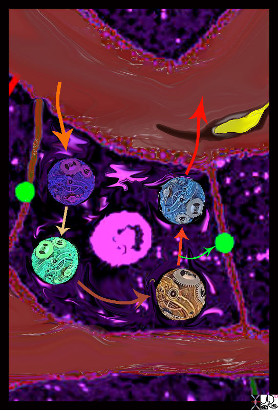
Receiving Processing Exporting Optimal Spatial Relationships Necessary |
| When the metabolic products enter the cell (orange arrow) they also have to enter a factory line that has a different arrangement than liver cord/sinusoid arrangement of the liver lobule. The organelles in the cell have to be spatially positioned and connected for the metabolic and biochemical processes to take place. The exact mechanisms and organelles responsible for the processwill be discused in detail in future modules and are represented here as organelles with cogs situated within the cell cytoplasm. The metabolic substrates enter the cell (light orange arrow) and are processed by the dark blue organelle, then exported to the light green organelle and so on, until the final step where a by product called bile (lime green arrow) is transported to the bile canaliculus ( green sphere between two cells) and the primary product is exported (red arrow) into the portal circulation (maroon).
13062b.4k08.8 Davidoff MD D avidoff art
|
The physical location of structures described above allows the component parts to hand off metabolic products from one to the other, as one might imagine happens in the factory line typified for example in production of a motor car. This was one of Henry Fords contributions to our factory industry – the factory line where each worker performed his or her repetitive function and then passed on the product to the next in line, who, in turn adds another component, screws, and tightens. As the product is passed on in the line, it evolves in an efficient manner mostly because of the spatial arrangement of the workers. It is almost as if Henry Ford understood liver structure and function when he came up with the idea. Biology has perfected these processes over thousands of years and has a lot more to teach us if we could only look, comprehend, and apply them to our macroscopic world.
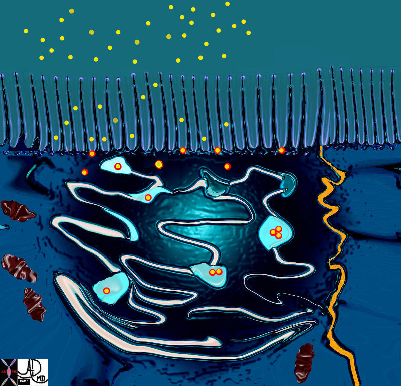
When a chemical message needs to be communicated, the spatial relationship between the giver and receiver has to be optimised . Examples include messages between two nerves, or between a nerve and a muscle, between a hormone and a receptor, or between a drug and a receptor. The diagram of the synapse below, shows the spatial relationship between the afferent (presynaptic) nerve and the efferent (postsynaptic) nerve. In this instance the two cells at the site of the synapse are quite different in their configuration. The presynaptic nerve is designed to optimise the release of the chemical message, the postsynaptic is designed and positioned to receive the chemical and its message. The space between them has to be appropriately sized and positioned to enable the message to pass through.
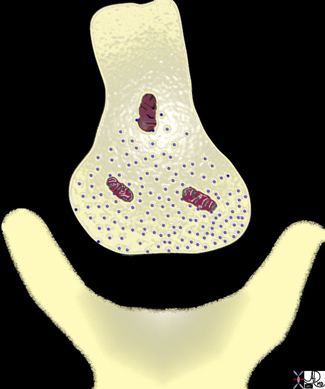
Synaptic Cleft Requiring Chemical Transmitters eg Acetylcholine to Transmit Impulse Across Nerve Junctions |
| 72046.800 mitochondria transmitter vesicles presynaptic terminal post synaptic terminal soma of neuron synaptic cleft acetyl choline norepinephrine dopamine serotonin forces chemical energy function principles Davidoff art Davidoff drawing Davidoff MD |
Body Spaces
At a macroscpic level the body houses the organs in cavities and spaces
Cavities of the body
Cranial Cavity
Thoracic Cavity
Abdominal Cavity
Pelvic Cavity
Cranial Cavity
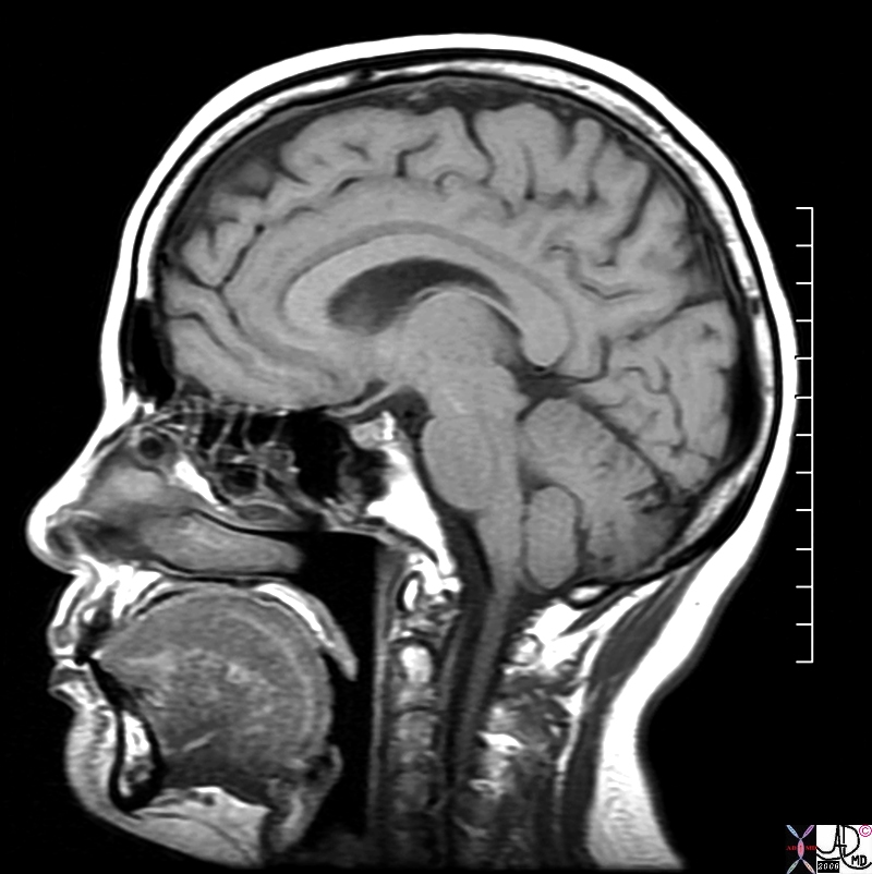
The cranial cavity, is the space formed inside the bony skull that houses the brain. The capacity of an adult human cranial cavity is 1,200-1,700ccs. The cranial cavity in the adult is a non compliant vault – that fits the brain prerfectly with little wiggle room. Aside from the small cushion effect of the meninges and surrounding cerebrospinal fluid the brain comes into close physical contact with the vault. As a relatively soft structure the brain requires this spatial arrangement to protect it.
Cranial Cavity Housing the Brain |
| The cranial cavity in the adult is a non compliant vault – that fits the brain prerfectly with little wiggle room. The MRI above is a sagittal projection, and shows the small space between the brain and the skull that is filled with memninges and cerebrospinal fluid.
71053.800 brain normal skull cranial cavity spinal cord cerebellum MRI T1 weighted normal anatomy Courtesy Ashley Davidoff MD copyright 2008 |
The Chest Cavity
The chest cavity or thoracic cavity, is the space formed inside the rib cage that houses the heart, lungs and mediastinum. It is connected to the neck at the thoracic inlet, and separated from the abdominal cavity by the diaphragm. The major organs that lie within the chest cavity include the heart with great vessels, lungs with airways, and the esophagus. The spinal cord lies posteriorly in its own chamber within the vertebral column and is not traditionally considered part of the thoracic cavity.
The lungs with pleura and airways take up the largest space. The lung at full inspiration can take up to 6 litres of air.
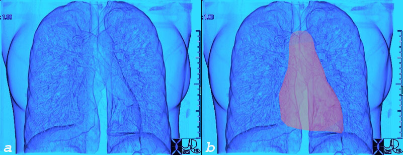
The Lungs in the Chest |
|
The lungs take up the largest space in the chest. They are paired and surround the heart (pink) which lies in the centre of the chest and occupies less space. 45770b02c.jpg chest lungs fx normal chest CT anatomy Courtesy Ashley Davidoff MD Davidoff |
The lungs surround the heart and are surrounded by by two layers of pleura that are adhered together by a thin layer of fluid.
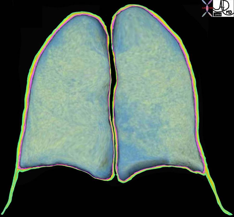 
Spaces in the Chest |
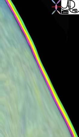
| The teal green lungs are surrounded by the visceral pleura (pink) pleural space (yellow) and parietal pleura (green) as seen in the both the image on the left and the zoomed image of the layers on the right.
32634b10 32634b11b01 The surface of the lung is magnified in the following two images. Fig 1a shows the pink visceral pleura, the yellow pleural space, and the green parietal pleura. Fig 1b shows the costophrenic sulcus where the visceral pleura is absent and two layers of parietal pleura face each other. Courtesy Ashley Davidoff MD . 32634b11b01 32634b11b |
When we inspire, the muscles of the rib cage move out, pull on the outside layer of the pleura, (parietal pleura) which in turn pulls on the inner layer of visceral pleura, which in turn pulls on the lungs, which then expand and pull in fresh air. The fluid layer in between the two layers of pleura enable the two smooth layers of pleura to adhere together by capillary force in the same way that two wet pieces of smooth glass are inseparable by virtue of the same capillary forces.
Abdominal Cavity
The abdominal cavity, is the space formed by a diaphragm of muscle above and a diaphragm of pelvic muscle below, and usually includes the pelvic cavity. It mostly houses organs of the gastrointestinal system, genitourinary system, but components of the cardiovascular system, nervous system, endocrine system, reticuloendothelial system, and musculoskeletal system are present to enable the abdominal cavity to connect with the thorax and all the other parts of ther body.
The major organs that lie within the abdominal cavity include the liver, spleen, adrenals, kidneys, pancreas, gastrointestinal tract, uterus ovaries and prostate and bladder.
Unlike the cranial cavity and the thoracic cavity, there is little bony protection. Most of the protection comes from more elastic and pliable abdominal musculature.
The spinal cord lies posteriorly in its own chamber within the vertebral column and is not traditionally considered part of the abdominal cavity.
The distinction and border between the abdominal cavity proper and the pelvic cavity is ill defined and vague, and for the sake of discussion it is considered in this text as both part of the abdominal cavity, though a specific subsection of this text will discuss the pelvic cavity separately as well.
Spaces in the Abdomen
The abdominal cavity is divided into the peritoneal cavity and the retroperitoneal cavity. Each of these is further divided and the detail will be expanded in later modules. The peritoneal cavity is the larger of the two and it contains almost all the abdominal organs. The reroperitoneal space contains only the kidneys, ureters, the larger tributaries of the neurovascular system like the aorta, inferior vena cava, nerves and lymphatic duct.
Pelvic Cavity
The pelvic cavity, is the space formed by a diaphragm of pelvic muscle below and an open roof above where the distinction between the pelvic cavity and abdominal cavity is vague but best defined by the region of the bony pelvis, including the iliac crests and lumbosacral joint and sacral promontory and the pubic symphisis. It houses the uterus, ovaries and Fallopian tubes, the bladder and distal ureters, prostate vasa deferentia, sigmoid colon and rectum. Components of the cardiovascular system, nervous system, endocrine system, reticuloendothelial system, and musculoskeletal system are present to enable the pelvic cavity to connect with all the other parts of ther body.
Similar to the cranial cavity and the thoracic cavity, there is prominent bony protection by the pelvic bones.
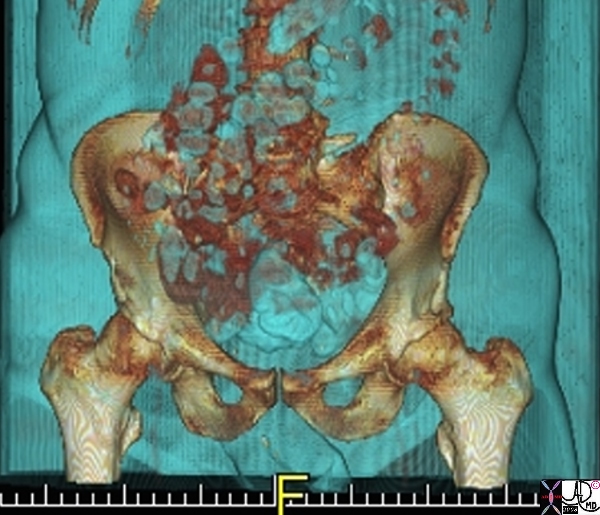
The Normal Pelvis |
|
The bony pelvis is shown in the above CT scan reconstruction. While the iliac crests and ischia and pubic symphisis are well shown, the sacral component is hidden by parts of the gastrointestinal tract. 71198b01 pelvis bone iliac crest pubic symphisis hip ischial tuberosity pubic bone normal anatomy CTscan 3D Courtesy Ashley Davidoff MD copyright 2008 |
Spaces in the Pelvis
Most of the pelvic cavity is part of the peritoneal cavity but the retroperitoneum does extend down into the pelvic cavity and thre is also a space called the extraperitoneal space.
Other Spaces
There are other spaces and compartments in the body that do not fit in to the confines of the larger spaces described above. These include the space of the spinal column, prevertebral spaces, compartments of the muscles bones and joints, the oral cavity, spaces of the neck, the vagina, the external space occupied by the penis and testes, and of course all the microscopic spaces of the tissues, cells and molecules that have a world of their own.
From the Cells to the Cities – Space – a Universal Principle |
|
The artists reconstruction of the space of cells and the the reality of the photograph of space in a seaside town in Cape Town South Africa, are seemingly different, but in reality the same with the universal principle being relevant to both of 57927.800 houses town organization position efficiency Muizenberg Cape South Africa Davidoff photography |
When one extends the concept of space beyond the body and into homes and towns, cities and countries and then of course planets and the universe – the universality of the principles unfold. When all is in order – and space is well used, and apportioned then peace and harmony exist. When on the other hand aggressive space occupation occurs the stories of disorder, disease, death, and misery are told in our history.
Forces
Many factors in the natural environment are prone to flux, such as temperature, UV radiation levels, and humidity, all of which challenge the ability of each organism to adapt to dynamic surroundings. However, other forces, such as gravity and electrostatic repulsion and attraction, are enacted upon and within the cells and molecules of the body, posing further obstacles to maintaining proper function. In this way, variable earthly forces create a minute-to-minute challenge for the organism and expose it to the possibility of damage and dysfunction.
Interactions
The particles of the universe are in constant motion – tumbling spinning and interacting in both chatic and in orderly fashions. The speed with which they move about relates to the energy they have – and is directly related to the heat that they contain. As they move about they interact with one another based on forces in the environment and forces that are innate. The forces may be attractive or repulsive in nature. The electrical forces of negative ands positive make the most intuitive sense. Opposite electrical forces attract and similar forces repel.
Biological units continually interact with each other and with the environment in a way that promotes either increased or decreased order within an organism. Waves, particles, and cells may all collide and interact in a continuous exchange of energy, as depicted by the image below.
Links and Connections at a Subcellular Level Intestinal Epithelial Cell – Golgi Apparatus and Intercellular Borders |
| 71798b13b03.800 cell endoplasmic reticulum mitochondria fat droplet absorbtion by microvilli processing cisterna interdigitaion of cells pinocytic vesicle intercellular space function absorbtion Davidoff art Davidoff MD |
States of Being
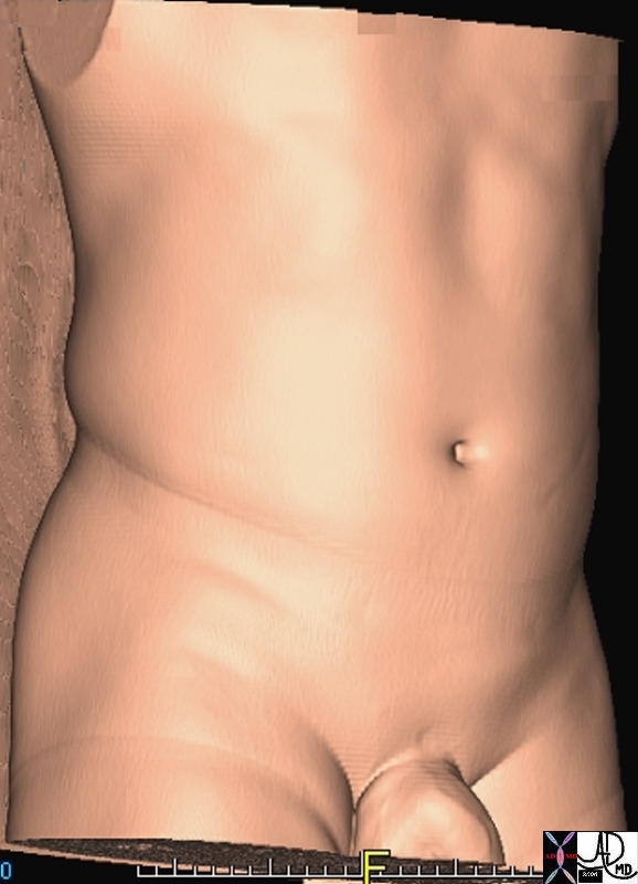
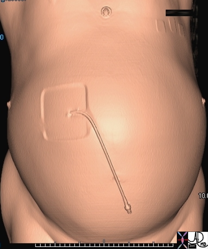
Within the universe, more particularly, our world, there are states of both order and disorder, which are relative to each other and govern the function of cells and the body. For instance, when a cell or organ experiences trauma its disorder increases relative to its order, leading to compromised function, disease, and, in some cases, death. The images below of a healthy abdomen versus one that is distended due to liver disease exemplify this.
|
Health and Disease of the Abdomen |
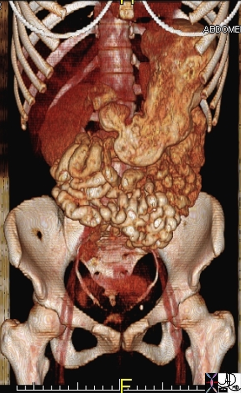
|
The image on the left repesents a normal male abdomen reconstructed from a CTscan. The image on the right is also from a male adult patient who has liver cirrhosis and as a result has accumulated a transudate in the abdominal cavity. This is called ascites and causes the abdomen to bulge. The small catheter is used to remove the fluid. Courtesy Ashley Davidoff MD |
Health and Disease
Health and disease represent the proper and improper function of the human body, respectively. In other words, they are conditions that result from the relative states of order and disorder described above, where disorder within a cell or the body corresponds to disease and malfunction.
Function of the Physician
The function of a physician is to provide compassionate care using prudence and knowledge in the diagnosis and treatment of disease. Underlying the interpersonal relationship the clinician has to be versed in the depth and breadth of the knowledge base and has to be able to be prudent in judgement in the application of this knowledge. Given these responsibilities the process of becoming a doctor is difficult and requires years of dedication and study. It is my hope however, to aid in the progression from college to medical school by presenting prospective medical students with a preview of the vast medical knowledge that awaits them should they pursue a career in medicine. Indeed, the principles I have outlined here are representative of those imparted in the first years of medical school and by presenting them in this format I seek to promote early understanding of medicine in physicians-to-be and provide them with a context in which to understand the detail to come in medical school.